Imaging Services
Stay connected to your care with MyChart® by Hawai‘i Pacific Health – message your provider, view test results, complete forms, and more.
Service Overview
Imaging is an important diagnostic tool that is used by many medical specialties.
Our imaging staff is expertly trained and certified to provide you with accurate, efficient imaging services in a comfortable, supportive environment.
Conditions & Treatments
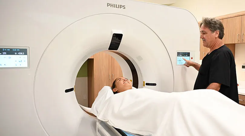
Medical imaging services are diagnostic procedures. Technology, like X-rays, magnetic fields, or sound waves, create images of the inside of the body. These images help doctors effectively diagnose injuries, confirm diseases, and monitor treatments.
Common types of imaging include MRIs, CT scans, ultrasounds, and mammography.
- Ultrafast/Electron Beam CT Scan
- Bone Scans
- Mammograms
- Nuclear Medicine
- Positron Emission Tomography (PET)
- Chest X-Ray
- Abdominal X-Rays
- X-rays of the Skull
- CT Screening Boosts Lung Cancer Survival
Interventional Radiology
Angiography
Angiography is an X-ray examination of the blood vessels that allows doctors to see how blood circulates within the body. It is used when a blockage in the flow of blood or abnormality of a blood vessel is suspected.
CT Scan
Advanced imaging technology can better detect diseases at an early stage, when a wider array of effective treatment options may be available.
MRI
An MRI, or Magnetic Resonance Imaging, is a test using strong magnetic fields and radio waves to produce detailed images of organs and tissues.
Care at Kapiʻolani
Kapiʻolani Medical Center for Women & Children is Hawaiʻi’s leader in pediatric and women’s health. Our Imaging Department specializes in diagnostic services for newborns, children and teens, while also providing general imaging for select adult studies. We combine state-of-the-art technology with a compassionate, family centered approach to ensure accurate results in a safe, supportive environment.
Our department provides a full range of imaging modalities:
- X‑Ray (Radiography): Quick, low‑dose exams that capture detailed images of bones, chest, and other body structures. We are uniquely equipped to provide specialty pediatric imaging.
- Fluoroscopy: Real‑time imaging that allows physicians to observe movement inside the body, such as swallowing studies or gastrointestinal exams.
- CT (Computed Tomography): Advanced cross sectional imaging. We use pediatric‑specific protocols to minimize radiation exposure while ensuring clear results.
- Ultrasound: Safe, radiation free imaging for soft tissues and organs.
- Echocardiography (Echo): Specialized ultrasound of the heart.
- MRI (Magnetic Resonance Imaging): Detailed imaging using magnetic fields, with child friendly protocols and sedation options when needed. Our staff are highly specialized in pediatric and breast MRI.
- Nuclear Medicine: Functional imaging using small amounts of radiotracers to evaluate organ function and detect disease.
Care at Pali Momi
Your Destination for Advanced Imaging and Women’s Health in Central and West O‘ahu
Pali Momi Medical Center is a leader in innovation and patient-centered care. As the first in West Oʻahu to introduce a permanent MRI, a 320-slice CT scanner, and a cardiac catheterization program, we continue to set the standard for advanced diagnostic and interventional services. We offer state-of-the-art technology for accurate, timely diagnosis, maintain radiation doses well below national standards for patient safety, and provide convenient scheduling—including early mornings and weekends—all within a comfortable, patient-focused environment.
Comprehensive Imaging Services
- CT Imaging – We provide a full range of CT services, including CT Lung Screening, Cardiac CT, and stroke imaging as part of our Stroke Care Center designation. Our team also performs CT-guided biopsies along with general CT studies, ensuring precision and timely diagnosis for complex conditions.
- Diagnostic Radiology - Our highly skilled technologists, trained to Trauma II standards, work closely with our expert radiologists to deliver precise, high-quality imaging across a wide range of conditions. This collaboration supports specialized care, including our Bone and Joint Center, ensuring accurate diagnosis and treatment planning. We also provide fluoroscopy for dynamic imaging studies, offering real-time visualization to guide complex procedures safely and effectively.
- MRI Services – Including 1.5T and 3T MRI systems, we deliver superior image clarity for complex exams such as comprehensive breast MRI and high-resolution prostate MRI studies—ensuring accurate diagnosis and treatment planning. To enhance patient safety, we have an MRI Safety Officer on staff who coordinates care for patients with metal implants, providing expert guidance and peace of mind.
- Nuclear Medicine Diagnostics – Our team of nuclear medicine technologists and radiologists carefully evaluates each case to determine the most appropriate diagnostic study or specialized treatment. This collaborative approach ensures accuracy and personalized care. Services include Cardiac Stress Tests, Myocardial Perfusion Imaging, Gastric Emptying Studies, HIDA Scans, Bone Scans, Lung Scans, GI Bleed Studies, Renal Scans, Lymphoscintigraphy, and Thyroid Uptake Exams—all designed to provide precise answers for complex conditions.
• Ultrasound – Our skilled team—holding multiple advanced certifications—provides high-quality imaging for general, abdominal, renal, thyroid, and vascular studies. We also perform ultrasound-guided biopsies in collaboration with radiologists and offer UroNav prostate biopsy, delivering targeted precision for prostate care.
• Interventional Radiology (IR) - Our dedicated IR Suite offers minimally invasive, image-guided procedures for vascular, oncologic, and other targeted therapies—designed to reduce recovery time and improve outcomes. Additionally, our IR services include prostate and uterine embolization’s, nephrostomy tube placement, gastrostomy, biliary drainage, and biliary rendezvous procedures.
• Pali Momi Women’s Center - Conveniently located near Pearlridge Center, the Pali Momi Women’s Center offers a spa-like environment dedicated to women’s health. Services include 3D mammography, breast ultrasound and non-surgical biopsy, and a comprehensive Bone Health Program with DEXA scanning. Patients also benefit from dedicated navigators and cancer support programs, ensuring compassionate care every step of the way.
• Radiologists - Our team of radiology physicians combines fellowship-trained interventional specialists with subspecialists dedicated to precision and innovation. From minimally invasive procedures to advanced neurological imaging, we provide care that improves outcomes. With expertise in breast and prostate imaging, orthopedic diagnostics, and nuclear medicine, we use state-of-the-art technology to support early detection and personalized treatment. Our radiologists bring decades of experience to every image, delivering clarity and confidence in every diagnosis. We don’t just interpret—we uphold the highest standards in medical imaging.
Experience advanced imaging and women’s health care with compassion and expertise.
Care at Straub Benioff
Straub Benioff Medical Center Imaging Services: The Most Advanced Care with Compassionate Expertise
At Straub Benioff Medical Center, we provide a full spectrum of imaging services from routine X-rays to the most advanced interventional radiology procedures all designed to deliver answers with precision and care.
- General X-rays: Produces high quality X-rays to support all providers especially the Bone and Joint Center.
- MRI Excellence: Our new 1.5T MRI system coupled with our highly skilled staff safely accommodates nearly every type of exam that isn’t contraindicated, offering clear, detailed images to guide diagnosis and treatment.
- Comprehensive CT Imaging: Our CT technologists specialize in complex exams across cardiology, orthopedics, ENT, neurology, gastrointestinal, and genitourinary specialties.
- General Ultrasound Expertise: Safe and radiation-free, ultrasound helps providers evaluate organs such as the liver, kidneys, gallbladder, and thyroid with ease and accuracy.
- Complex Vascular Lab Imaging: Using painless ultrasound, our technologists assess blood flow in veins and arteries to detect blockages or narrowing all without radiation or needles.
- Women’s Services: We offer digital mammography, tomosynthesis, breast ultrasound, MRI breast exams, and DEXA (bone density) scans. Straub Benioff proudly features the only state’s upright stereotactic breast biopsy system, allowing patients to undergo biopsies in a comfortable seated position.
- Nuclear Medicine Innovation: Our Nuclear Medicine Department provides advanced radioligand therapies such as Pluvicto, Lutathera, and Xofigo, alongside diagnostic studies including HIDA scans, gastric emptying exams, and cardiac stress tests.
- Interventional Radiology: Our team performs the widest range of vascular and non-vascular procedures within the system including but not limited to cerebral angiography, aneurysm coiling, embolization, nephrostomy tube placement, gastrostomy, biliary drainage, biliary rendezvous, and TIPS procedures.
- Expert Radiologists: More than 10 fellowship-trained radiologists bring subspecialty expertise in neuroradiology, musculoskeletal imaging, cardiac imaging, breast imaging, interventional radiology, and body imaging.
Straub Benioff combines cutting-edge technology with compassionate care ensuring every patient receives the answers they need, delivered by experts they can trust.
Care at Wilcox
The center is equipped with state-of-the-art technology to serve patients across the inpatient, outpatient, and emergency settings.
The spectral-detector CT scanner produces comprehensive images for patients of all ages, including children, and with a range of medical needs, such as bariatrics. The machine generates high-quality, exceptionally clear images with faster setup times and shorter exam times. This results in a lower dose of radiation as well as a more comfortable overall experience.
The 320-slice CT scanner offers advanced imagery full-body scanning in a single rotation of the X-ray tube. This faster scan time is helpful for patients who have a hard time staying still for extended periods.
Advanced features of the CT scanners at Wilcox help medical teams detect, diagnose and monitor both common and chronic diseases, disorders and conditions, including cancer, heart disease and bone fractures.
Imaging services are available 24 hours a day, seven days a week. Other services offered include X-ray; Ultrasound; Mammography; MRI; Nuclear Medicine and limited interventional radiologic procedures. Board-certified physician radiologists provide quality care and timely interpretation of radiographic procedures.
Services are by referral from a patient’s primary care physician, who will use these internal images of the body to assist with diagnosis and treatment.
Our Care Locations
Oʻahu
Kapiʻolani Medical Center for Women & Children Kapiʻolani Medical Center for Women & Children
1319 Punahou St.
Honolulu, HI 96826
Phone: 808-983-6000
Get directions
Details
Fetal Diagnostic Center
Fetal Diagnostic Center
Kapi‘olani Medical Center for Women & Children, Physician Office Tower, Suite 540
1319 Punahou St.
Honolulu, HI 96826
Phone: 808-983-8559
Get directions
Details
Pali Momi Medical Center
Kapiʻolani Medical Center for Women & Children
1319 Punahou St.
Honolulu, HI 96826
Phone: 808-983-6000
Get directions
Details
Fetal Diagnostic Center
Fetal Diagnostic Center
Kapi‘olani Medical Center for Women & Children, Physician Office Tower, Suite 540
1319 Punahou St.
Honolulu, HI 96826
Phone: 808-983-8559
Get directions
Details
Pali Momi Medical Center
 Pali Momi Medical Center
98-1079 Moanalua Road
‘Aiea, HI 96701
Phone: 808-486-6000
Get directions
Details
Pali Momi Outpatient Center
Pali Momi Medical Center
98-1079 Moanalua Road
‘Aiea, HI 96701
Phone: 808-486-6000
Get directions
Details
Pali Momi Outpatient Center
 Pali Momi Outpatient Center
98-1005 Moanalua Road
ʻAiea, HI 96701
Phone: 808-486-6000
Get directions
Details
Pali Momi Women's Center
Pali Momi Outpatient Center
98-1005 Moanalua Road
ʻAiea, HI 96701
Phone: 808-486-6000
Get directions
Details
Pali Momi Women's Center
 Pali Momi Women's Center
98-1005 Moanalua Road
ʻAiea, HI 96701
Phone: 808-485-4500
Get directions
Details
Straub Benioff Medical Center
Pali Momi Women's Center
98-1005 Moanalua Road
ʻAiea, HI 96701
Phone: 808-485-4500
Get directions
Details
Straub Benioff Medical Center
 Straub Benioff Medical Center
888 S. King St.
Honolulu, HI 96813
Phone: 808-522-4000
Get directions
Details
Straub Benioff Medical Center – Doctors on Call at Sheraton Waikīkī
Straub Benioff Medical Center – Doctors on Call at Sheraton Waikīkī
2255 Kalākaua Ave.
Manor Wing, Lower Level
Honolulu, HI 96815
Phone: 808-971-6000
Get directions
Details
Straub Benioff Medical Center – Hawaiʻi Kai Clinic
Straub Benioff Medical Center – Hawaiʻi Kai Clinic
Koko Marina Shopping Center
7192 Kalanianaʻole Highway, Suite A200
Honolulu, HI 96825
Phone: 808-396-6321
Get directions
Details
Straub Benioff Medical Center – Kāhala Clinic & Urgent Care
Straub Benioff Medical Center
888 S. King St.
Honolulu, HI 96813
Phone: 808-522-4000
Get directions
Details
Straub Benioff Medical Center – Doctors on Call at Sheraton Waikīkī
Straub Benioff Medical Center – Doctors on Call at Sheraton Waikīkī
2255 Kalākaua Ave.
Manor Wing, Lower Level
Honolulu, HI 96815
Phone: 808-971-6000
Get directions
Details
Straub Benioff Medical Center – Hawaiʻi Kai Clinic
Straub Benioff Medical Center – Hawaiʻi Kai Clinic
Koko Marina Shopping Center
7192 Kalanianaʻole Highway, Suite A200
Honolulu, HI 96825
Phone: 808-396-6321
Get directions
Details
Straub Benioff Medical Center – Kāhala Clinic & Urgent Care
 Straub Benioff Medical Center – Kāhala Clinic & Urgent Care
Kūʻono Marketplace
4210 Waiʻalae Ave., Suite 501
Honolulu, HI 96816
Phone: 808-462-5300
Get directions
Details
Straub Benioff Medical Center – Kapolei Clinic & Urgent Care
Straub Benioff Medical Center – Kapolei Clinic & Urgent Care
Ka Makana Ali‘i
91-5431 Kapolei Parkway, Suite 1706
Kapolei, HI 96707
Phone: 808-426-9300
Get directions
Details
Straub Benioff Medical Center – King Street Clinic
Straub Benioff Medical Center – King Street Clinic
888 S. King St.
Strode Building, 1st floor
Honolulu, HI 96813
Phone: 808-522-4000
Get directions
Details
Straub Benioff Medical Center – Mililani Clinic & Urgent Care
Straub Benioff Medical Center – Kāhala Clinic & Urgent Care
Kūʻono Marketplace
4210 Waiʻalae Ave., Suite 501
Honolulu, HI 96816
Phone: 808-462-5300
Get directions
Details
Straub Benioff Medical Center – Kapolei Clinic & Urgent Care
Straub Benioff Medical Center – Kapolei Clinic & Urgent Care
Ka Makana Ali‘i
91-5431 Kapolei Parkway, Suite 1706
Kapolei, HI 96707
Phone: 808-426-9300
Get directions
Details
Straub Benioff Medical Center – King Street Clinic
Straub Benioff Medical Center – King Street Clinic
888 S. King St.
Strode Building, 1st floor
Honolulu, HI 96813
Phone: 808-522-4000
Get directions
Details
Straub Benioff Medical Center – Mililani Clinic & Urgent Care
 Straub Benioff Medical Center – Mililani Clinic & Urgent Care
Town Center of Mililani
95-1249 Meheʻula Parkway, Building M
Mililani, HI 96789
Phone: 808-625-6444
Get directions
Details
Straub Benioff Medical Center – Pearlridge Clinic
Straub Benioff Medical Center – Pearlridge Clinic
98-151 Pali Momi St.
Suite 142
‘Aiea, HI 96701
Phone: 808-483-6400
Get directions
Details
Straub Benioff Medical Center – Ward Village Clinic & Urgent Care
Straub Benioff Medical Center – Ward Village Clinic & Urgent Care
1001 Queen St., Suite 102
Honolulu, HI 96814
Phone: 808-462-5200
Get directions
Details
Straub Benioff Medical Center – Mililani Clinic & Urgent Care
Town Center of Mililani
95-1249 Meheʻula Parkway, Building M
Mililani, HI 96789
Phone: 808-625-6444
Get directions
Details
Straub Benioff Medical Center – Pearlridge Clinic
Straub Benioff Medical Center – Pearlridge Clinic
98-151 Pali Momi St.
Suite 142
‘Aiea, HI 96701
Phone: 808-483-6400
Get directions
Details
Straub Benioff Medical Center – Ward Village Clinic & Urgent Care
Straub Benioff Medical Center – Ward Village Clinic & Urgent Care
1001 Queen St., Suite 102
Honolulu, HI 96814
Phone: 808-462-5200
Get directions
Details
Kauaʻi
Wilcox Medical Center Wilcox Medical Center
3-3420 Kūhiō Highway
Līhu‘e, HI 96766
Phone: 808-245-1100
Get directions
Details
Kauaʻi Urgent Care
Wilcox Medical Center
3-3420 Kūhiō Highway
Līhu‘e, HI 96766
Phone: 808-245-1100
Get directions
Details
Kauaʻi Urgent Care
 Kauaʻi Urgent Care
4484 Paheʻe St.
Līhuʻe, HI 96766
Phone: 808-245-1532
Get directions
Details
Kauaʻi Urgent Care
4484 Paheʻe St.
Līhuʻe, HI 96766
Phone: 808-245-1532
Get directions
Details